
The muscles used for mastication (chewing) include:
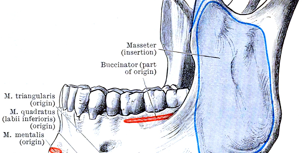
1. Masseter: One of the strongest muscles in the body, the masseter elevates the mandible (jawbone) to close the mouth.
2. Temporalis: A fan-shaped muscle located on the side of the head, the temporalis elevates and retracts the mandible.
3. Medial Pterygoid: This muscle works in tandem with the masseter to elevate the mandible. It also helps with side-to-side movement of the jaw.
4. Lateral Pterygoid: This muscle is responsible for protruding the mandible (pushing it forward) and assisting in opening the mouth. It also facilitates side-to-side movements.

The muscles of mastication work together in a coordinated manner to achieve various movements of the jaw, allowing for effective chewing, biting, and grinding of food.
Here’s how they function collectively:
1. Elevation of the Mandible (Closing the Mouth):
- Masseter: Contracts to elevate the mandible, bringing the teeth together.
- Temporalis: Contracts to elevate and retract the mandible, assisting in closing the mouth and pulling the jaw backward.
- Medial Pterygoid: Contracts to elevate the mandible, working in synergy with the masseter.
2. Depression of the Mandible (Opening the Mouth):
- Lateral Pterygoid: Contracts to pull the mandible forward and downward, helping to open the mouth. Other muscles such as the digastric, mylohyoid, and geniohyoid also assist in this movement.
3. Protrusion of the Mandible (Pushing the Jaw Forward):
- Lateral Pterygoid: Contracts to pull the mandible forward.
- Medial Pterygoid: Assists in protruding the mandible.
4. Retraction of the Mandible (Pulling the Jaw Backward):
- Temporalis: The posterior fibers of the temporalis muscle contract to retract the mandible.
5. Lateral Movements of the Mandible (Side-to-Side Movements):
- Lateral Pterygoid: When the lateral pterygoid muscles on one side contract, they pull the mandible towards the opposite side.
- Medial Pterygoid: Assists in the side-to-side movement by elevating the mandible on one side.
Coordinated Action
1. Chewing (Mastication):
During chewing, the jaw moves in a complex cycle involving elevation, depression, protrusion, retraction, and lateral movements. The masseter, temporalis, medial pterygoid, and lateral pterygoid muscles coordinate these actions:
- Food is initially bitten and cut by the incisors with the help of the masseter and temporalis muscles.
- As chewing continues, the lateral and medial pterygoid muscles facilitate grinding by moving the jaw side to side.
- The masseter and temporalis muscles maintain the force needed to keep the teeth together while grinding.
2. Grinding:
The lateral movements (mediated by the pterygoid muscles) are crucial for grinding food between the molars.
This intricate coordination ensures that food is broken down efficiently for digestion. The neural control from the trigeminal nerve (cranial nerve V), particularly the mandibular branch (V3), innervates these muscles, enabling precise and smooth movements.

The neural control of mastication involves a complex interaction between the central and peripheral nervous systems. Here's how this control is orchestrated:
Central Nervous System (CNS)
1. Cerebral Cortex:
- Motor Cortex: The primary motor cortex, located in the precentral gyrus of the frontal lobe, initiates voluntary movements, including those for mastication.
- Somatosensory Cortex: Provides feedback on the position of the jaw and the force being applied during chewing.
2. Brainstem:
- Pons: Contains the trigeminal motor nucleus, which is crucial for mastication.
- Medulla Oblongata: Involved in reflexive movements and coordination.
Peripheral Nervous System (PNS)
1. Trigeminal Nerve (Cranial Nerve V):
-
Mandibular Branch (V3): This branch of the trigeminal nerve is primarily responsible for innervating the muscles of mastication. It has both sensory and motor functions:
- Motor Component: Innervates the masseter, temporalis, medial pterygoid, and lateral pterygoid muscles.
- Sensory Component: Provides sensory feedback from the lower third of the face, the anterior two-thirds of the tongue (excluding taste), and the mucosa of the mouth.
Neural Pathways and Reflexes
1. Voluntary Control:
- Initiated in the motor cortex and transmitted via upper motor neurons through the corticobulbar tract to the trigeminal motor nucleus in the pons. From there, lower motor neurons carry the signal to the muscles of mastication.
2. Reflex Control:
- Jaw-Jerk Reflex: A monosynaptic reflex involving the sensory and motor components of the mandibular branch of the trigeminal nerve. Tapping the chin causes a quick contraction of the jaw-closing muscles, demonstrating the reflex arc.
- Chewing Reflex: Involves a more complex circuit with inputs from sensory receptors in the mouth and teeth, processed by the brainstem and higher brain centers to modulate the force and rhythm of chewing.
Sensory Feedback
1. Proprioception:
Sensory receptors in the muscles, tendons, and joints provide information about the position and movement of the jaw. This feedback is crucial for adjusting the force and timing of muscle contractions during chewing.
2. Mechanoreceptors:
Located in the periodontal ligament around the teeth, these receptors provide information about the texture and consistency of food, aiding in the modulation of chewing force.
Integration and Coordination
- The brainstem integrates sensory inputs and motor commands, ensuring smooth and coordinated movements of the jaw.
- Higher brain centers, including the cortex, can modulate these reflexes and voluntary movements based on sensory feedback and cognitive inputs, such as deciding to chew more thoroughly or to stop chewing.
Overall, the neural control of mastication is a highly coordinated process involving multiple brain regions and neural pathways to ensure efficient and effective chewing.
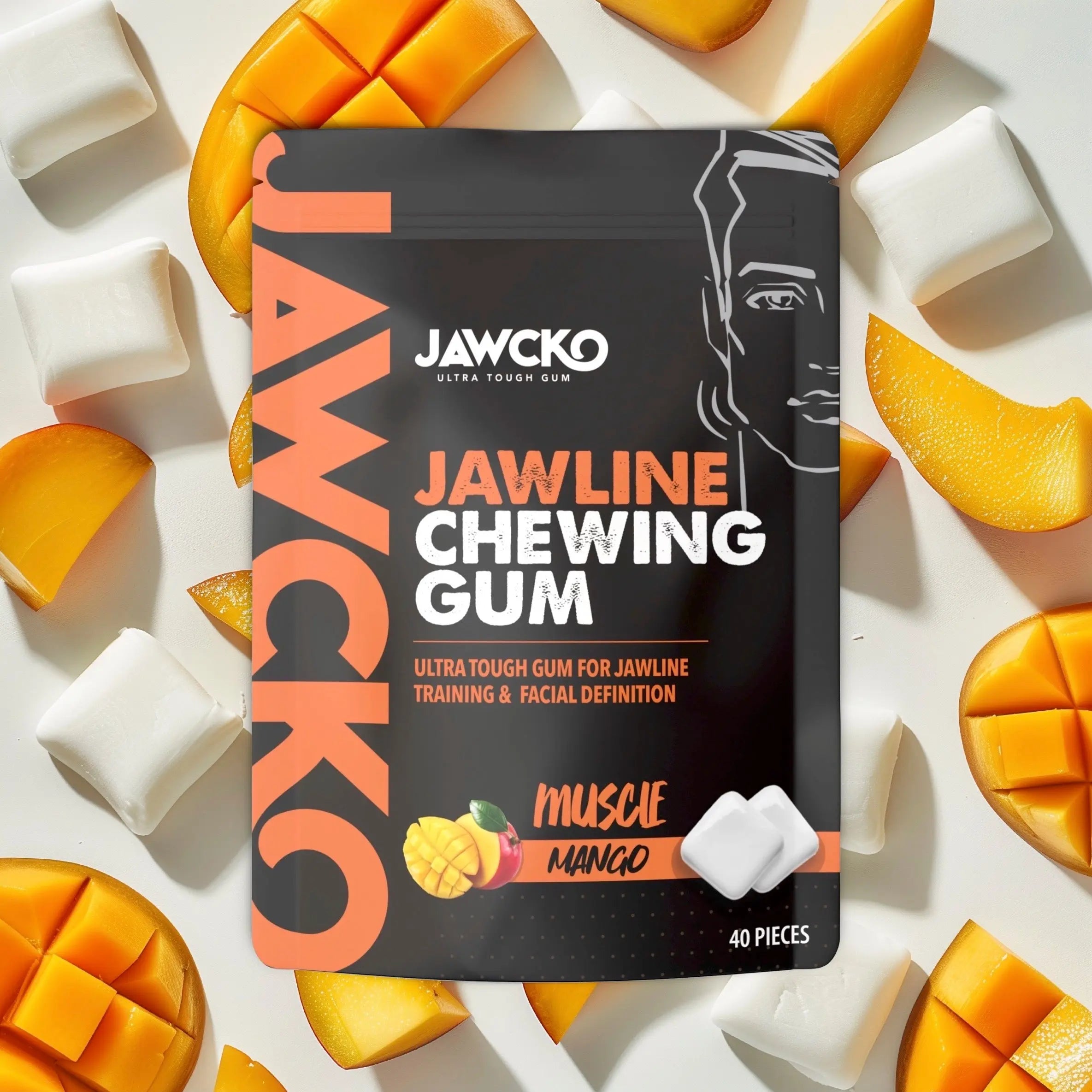
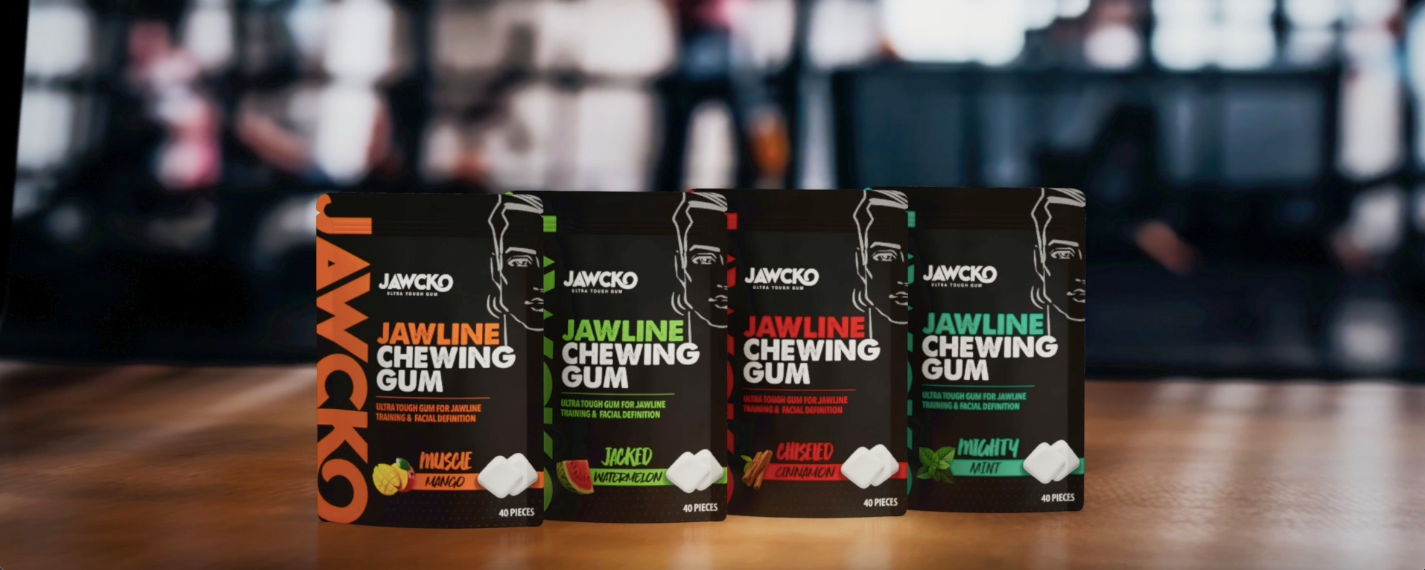
What This Means at JAWCKO
Mastication is a complex process that involves the seamless coordination of your brain, muscles, and psychology. Your brain controls the precise movements of the jaw through neuromuscular signals, activating key muscles like the masseter, temporalis, and pterygoids to generate force and endurance. Meanwhile, your psychology plays a role in habit formation, muscle engagement, and the mind-muscle connection, ensuring consistency and effort in jaw training. At JAWCKO, we are dedicated to understanding the intricate science behind jawline development, using cutting-edge research to formulate the most effective jawline gum. By optimizing resistance, durability, and jaw muscle activation, our gum is designed to maximize results in both aesthetics and function—helping you achieve a stronger, more defined jawline with every chew. Science drives our innovation, ensuring that JAWCKO delivers real, measurable benefits for your facial fitness journey